 A medical tool that looks like a handheld pen can detect cancer cells in just seconds and help surgeons to more accurately extract tumors, said researchers on Wednesday.
The tool works in real time, and its accuracy is at a minimum equal to removing a sample of tissue and having it sent to pathologists, said a team of researchers from the University of Texas.
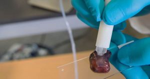
The pen uses just a drop of water to form an analysis and does not require any tissue to be cut. Researchers hope the pen can help make surgery less invasive and get all of the tumor while leaving behind as much of the healthy tissue possible.
She said that it is heartbreaking if that isn’t the case, but the new technology could improve odds that the surgeons can remove all trace of cancer during surgery.
The research team designed its pen and programmed the mass spectrometer so it could detect compounds that make thyroid, ovary, breast and lung tumors different from tissue that is healthy.
The researchers said the tool was accurate over 96% of the time. Another member of the research team said that any time a patient can be offered more precise, safer and quicker surgery, it is something we as researchers want to do.
The technology, he added, does all of that, as it allows the surgeon to be far more precise in the tissue that is removed and what is left behind.
Researchers tested the pen on over 250 human tumor samples, calibrating each type of tumor’s molecular signature. Tumor cells carry out different activity than do normal cells, and researchers have worked for decades using mass spectrometry in an attempt to identify the differences quickly.
The device is known as MasSpec Pen and enables automated and controlled delivery of a water droplet to the surface of the tissue for the efficient extraction of biomolecules the team explained.
At present, the technique most often carried out to diagnose cancer is through a pathologist who looks at the frozen sample of extracted tissue through a microscope. The job requires much skill, but can become time consuming and is known to produce errors.